What is Nail Intramedullari and How is it Used in Surgery?
The emergence of nail intramedullari has transformed surgical practices. Dr. Emily Carter, a renowned orthopedic surgeon, emphasizes its significance: "Nail Intramedullari is a game-changer for fracture stabilization.” This technique involves the insertion of a specialized rod into the bone marrow. It aligns and stabilizes fractured bones, promoting faster recovery.
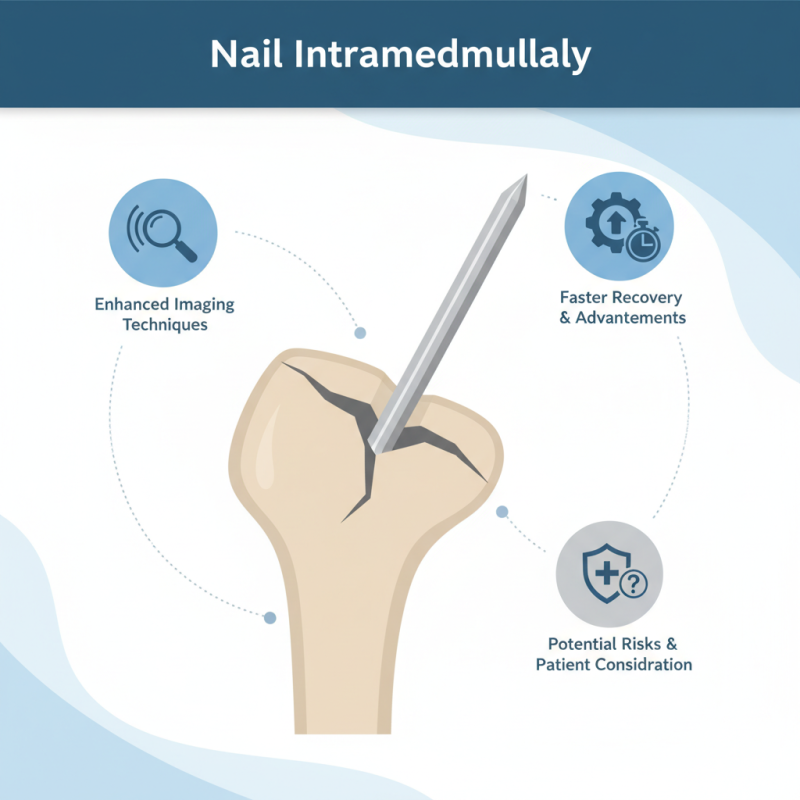
In recent years, advancements in technology have further refined the application of Nail Intramedullari. Surgeons now utilize enhanced imaging techniques for precise insertion. However, the procedure is not without challenges. Complications can arise from improper alignment or infection, creating hurdles in patient recovery. Reflection on these pitfalls is vital for continuous improvement.
Despite its benefits, some patients remain skeptical. Understanding the potential risks fosters informed decisions. As the field evolves, further research is essential to address concerns and refine techniques. Overall, Nail Intramedullari represents a significant advancement, though it requires thoughtful consideration by both surgeons and patients.
Definition and Overview of Intramedullary Nails in Orthopedic Surgery
Intramedullary nails are vital in orthopedic surgery, especially for treating fractures. They are metal rods inserted into the bone's medullary cavity. This method provides stable fixation, allowing for proper healing. According to recent reports, intramedullary nails significantly improve recovery times compared to traditional plating methods. Patients experience less pain and better alignment of bone fragments.
The design of intramedullary nails allows for minimal invasiveness. Surgeons often prefer them for femoral and tibial fractures. Approximately 80% of orthopedic surgeons rely on this technique for long bone stabilization. Despite their effectiveness, complications can arise. Surgeons must carefully consider potential risks, such as infection or improper alignment during insertion.
Tips: Always discuss surgical options thoroughly with your orthopedic surgeon. Understand the risks involved in using intramedullary nails. Research indicates that complications are higher in patients with underlying conditions. Staying informed empowers patients to make better choices about their care.
Nail Intramedullari Usage in Orthopedic Surgery
Historical Development of Intramedullary Nail Techniques and Innovations
Intramedullary nails have a long and fascinating history. Initially developed in the early 20th century, these devices offered a novel solution for bone fractures. Surgeons began to use these nails as a way to stabilize long bone fractures. The design has evolved significantly over the decades. Early versions were simple rods, but advancements have made them more effective and multifunctional.
Modern intramedullary nails include features such as locking mechanisms, which enhance stability. Innovations like minimally invasive techniques allow for quicker recovery times. These tools have transformed orthopedic surgery. They help in achieving better alignment and support for fractured bones. Yet, challenges remain. Complications can occur, and patient variability can affect outcomes. Surgeons must continuously learn and adapt.
**Tip:** Always stay informed about new techniques. Attend seminars and workshops to refine your skills.
Surgeons should also consider the individual needs of each patient. Every fracture is unique. A tailored approach can lead to better recovery. This continuous learning ensures effective treatment.
**Tip:** Collaborate with a multidisciplinary team for optimal patient care. Sharing insights can improve surgical outcomes.
Indications for the Use of Intramedullary Nails in Bone Fracture Treatment
Intramedullary nail fixation is a common method used in orthopedic surgery. It addresses various types of bone fractures effectively. Surgeons often recommend this technique for long bone fractures, particularly in the femur and tibia. The benefits include minimal soft tissue damage and stable internal support.
Using intramedullary nails can be indicated for specific types of fractures. For example, it is often chosen for diaphyseal fractures, where there is a risk of malunion. Additionally, it is suitable for fractures arising from accidents or high-impact sports. The placement of the nail aligns the bone fragments, promoting healing.
When considering intramedullary nails, here are some tips. Ensure a thorough medical evaluation first. This helps avoid complications during surgery. Recovery can vary, and sometimes, patients experience discomfort. Therefore, be prepared for a rehabilitation plan. Communicate openly with healthcare providers about your progress. This is important to address any concerns.
What is Nail Intramedullari and How is it Used in Surgery? - Indications for the Use of Intramedullary Nails in Bone Fracture Treatment
| Indication | Description | Common Types of Fractures | Benefits of Intramedullary Nails |
| Long Bone Fractures | Used for stabilizing fractures in long bones such as femur and tibia. | Femoral fractures, Tibial fractures | Provides stable fixation, allows early mobilization. |
| Complex Fractures | Used in cases where fractures are complicated by multiple fragments or instability. | Comminuted fractures | Improved alignment and reduced surgical time. |
| Non-union Fractures | Used in cases where a fracture has failed to heal correctly. | Delayed union, Non-union fractures | Enhances healing process and promotes bone regeneration. |
| Pathological Fractures | Used when fractures occur in bones weakened by disease. | Fractures in osteoporotic bones | Provides stability and minimizes further pain from instability. |
Surgical Procedures and Techniques Involving Intramedullary Nails
Intramedullary nailing is a common surgical technique for fixing fractures, especially in long bones like the femur and tibia. This method involves the use of a metal rod inserted into the medullary cavity. It enables stabilization of the fractured bone while allowing for early rehabilitation. According to a study published in the Journal of Orthopaedic Research, the use of intramedullary nails has shown a success rate of over 90% in certain types of fractures.
Surgical procedures using intramedullary nails require careful consideration. Surgeons must assess the fracture type and location before deciding on this technique. For instance, in unstable fractures, interlocking nails may provide additional stability. Reports indicate that complications can arise, such as postoperative pain and nail migration. In fact, a significant percentage of patients report some discomfort during recovery. Surgeons must weigh the benefits against these potential risks.
Proper technique is crucial in intramedullary nailing. Misalignment during the procedure can lead to deformities. Ensuring accurate placement of the nail is vital. Ideally, imaging guidance should be used to enhance accuracy. Additionally, ongoing training is important for surgical teams to stay updated on advancements in techniques and devices. Continuous reflection on outcomes and complications is essential for improving patient care in orthopaedic surgery.
Postoperative Care and Complications Associated with Intramedullary Nails
Intramedullary nailing is a surgical technique used for fractures, especially in long bones. It involves inserting a rod into the medullary cavity. While effective, there are postoperative challenges and complications to consider. According to recent data, complications occur in approximately 10-20% of intramedullary nail surgeries, which underscores the need for careful monitoring.
Postoperative care is crucial to mitigate these risks. Patients often experience pain and swelling. Regular assessments of limb function and alignment are vital. Infection is one of the most common complications. It can occur in up to 5% of cases, leading to prolonged recovery. Proper wound care and hygiene are essential in reducing this risk. Adjustments in mobility can help manage pain and ensure better healing.
Another concern is malunion or nonunion, where fractures do not heal correctly. Reports indicate that this happens in nearly 15% of cases. This situation often requires further surgical intervention. Active communication with healthcare providers can improve outcomes. Understanding these aspects can guide patients in their recovery journey.